Learn More
Leave A Message
Submit
Applications
Surface Reconstruction and Bulk Defect Engineering Stabilize Na-Layered Oxide Cathodes for High-Performance Sodium-Ion Batteries
A recent study led by Professor Yao Xiao's team at Wenzhou University reveals how surface reconstruction integrated with bulk defect engineering can overcome several long-standing stability limitations of Na-layered oxide cathodes. Using a combination of theoretical calculations, synchrotron X-ray absorption spectroscopy, resonant inelastic X-ray scattering, spherical-aberration-corrected scanning transmission electron microscopy, operando electrochemical mass spectrometry, and scanning electron microscopy, the researchers demonstrated that Y-induced bulk defect regulation and a robust reconstructed surface layer can work together to stabilize both bulk and interfacial chemistry. The strategy suppresses excessive lattice oxygen oxidation and O-O dimer formation, improves Na-ion transport, reduces transition-metal dissolution, relieves lattice strain, and enhances oxygen-redox reversibility. The result is significantly improved reaction kinetics, cycling stability, and electrochemical performance in high-voltage Na-layered oxide cathodes. The work, titled “Surface Reconstruction-Integrated Bulk Defect Engineering Beyond Conventional Chemical Modulation for Na-Layered Oxide Cathodes,” was published in Advanced Materials in 2026. Why Na-Layered Oxide Cathodes Need Better Stabilization The dual goals of reducing cost and extending service life continue to drive the development of energy-storage systems. Sodium-ion batteries are attracting increasing attention because sodium resources are abundant and relatively low in cost. Their operating mechanism is also similar to that of lithium-ion batteries, making them a promising route for large-scale energy storage. Among candidate cathode materials, transition-metal layered oxides with the general formula O3-type NaTMO2 are particularly attractive because of their relatively high theoretical capacity, straightforward synthesis, and relatively high tap density. However, deep desodiation at high voltage introduces a series of structural and interfacial problems. These can include phase transitions, transition-metal migration, lattice oxygen loss, and interfacial side reactions. Together, these processes can result in structural collapse, capacity decay, particle cracking, and pulverization. Conventional strategies such as surface coating, bulk doping, and oxygen-vacancy regulation can improve certain aspects of bulk, surface, or interfacial behavior, but it remains difficult to simultaneously optimize all three. To address these challenges, Professor Yao Xiao's team at Wenzhou University conducted an in-depth investigation using CIQTEK scanning electron microscopy as part of the multi-scale characterization workflow. With O3-NaNi1/3Fe1/3Mn1/3O2 as the model cathode, the researchers developed a surface reconstruction-integrated bulk defect engineering strategy to simultaneously regulate bulk, surface, and interfacial stability. By introducing an appropriate amount of Y into the material, the resea...
CIQTEK EPR200M with 3D-Printed Electrolytic Cell Enables Operando Detection of Radical Intermediates
Understanding the formation of radical intermediates is key to controlling electrochemical reaction rates and selectivity. These short-lived species at the electrode interface dictate outcomes, and relying solely on final products can lead to speculative mechanisms. With operando EPR using CIQTEK benchtop EPR200M, researchers can directly capture radicals in situ, mapping their formation sequence and structural fingerprints for robust mechanistic evidence. A recent collaboration between Beijing University of Technology (Sun Zaicheng / Liu Yichang), Tsinghua University (Yang Haijun), and Wuhan University (Lei Aiwen) introduced a novel 3D-printed electrolytic cell tailored for in situ EPR. Fabricated with high-precision digital light processing (DLP), this flat cell enables reproducible integration with electrochemical systems. Their results, published in Chemical Engineering Journal under the title Bespoke electrolytic cell for operando EPR tests: Revealing the formation and accurate structures of amino and phenolic radicals, demonstrate the workflow’s ability to uncover radical structures across representative reactions.   Methodological Breakthrough: 3D-Printed Flat Electrolytic Cell for Reproducible Operando EPR High-dielectric solvents commonly used in electrochemical cells reduce EPR signal-to-noise, making radical detection challenging. The flat cell design mitigates dielectric losses and enhances the resonator’s Q factor, improving operando EPR performance. Beyond physics, the cell is engineered for reproducibility. Using DLP 3D printing, electrode channels, positioning structures, and short-circuit protection are fixed during fabrication. This eliminates manual variability, reduces system resistance, and improves signal quality, while maintaining mechanical strength, solvent compatibility, and cost efficiency. This approach transforms operando EPR into a workflow of "standardized structural component + reproducible procedure", enabling cross-team and cross-system reproducibility and mechanistic comparison.   Time-Resolved Evidence Tracks Radical Formation in C–N Coupling In situ EPR with time-resolved acquisition allows mapping radicals in real-time, showing which species appear first and how they evolve. This provides a reproducible evidence chain at the intermediate level, moving mechanistic understanding beyond product-based inference.   Cycloaddition Intermediates Reveal Reaction Selectivity By comparing substrate-specific spectra and calculating spin density, EPR signals are directly translated into radical structural fingerprints. This forms a closed-loop framework for explaining regio- and chemo-selectivity in (3+2) cycloaddition reactions.   Solvent Effects Guide C–O Coupling Design In situ EPR shows that the same radical exhibits distinct spectra in MeCN versus HFIP. Combined with NMR, the study links solvent, radical structure, and reaction selectivity, providing an experimental evidenc...
CIQTEK SNVM Reveals Magnon Transport in BiFeO₃ (Advanced Functional Materials)
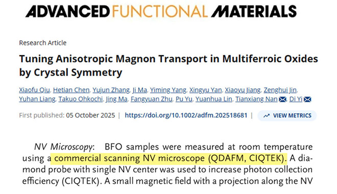
With the support of CIQTEK Scanning NV Microscopy (SNVM), researchers at Tsinghua University have directly visualized nanoscale spin cycloid structures in multiferroic BiFeO₃. This work, published in Advanced Functional Materials, provides the missing microscopic evidence linking crystal symmetry, magnetic structure, and anisotropic magnon transport, highlighting SNVM as a decisive tool for magnonics and low-power spintronic research.   The study used the CIQTEK Scanning NV Probe Microscope (SNVM) Research Background: Magnon Transport in Multiferroic Oxides Magnon-mediated spin currents can propagate in magnetically ordered insulators with nearly zero energy dissipation, making them highly attractive for next-generation low-power spintronic devices. In multiferroic materials such as BiFeO₃, the coupling between ferroelectric and antiferromagnetic orders enables electric field control of magnons, a long-standing goal in spintronics. Despite this promise, the microscopic origin of weakly anisotropic magnon transport in rhombohedral phase BiFeO₃, commonly referred to as R-BFO, has remained unresolved. Addressing this challenge requires direct real-space characterization of nanoscale magnetic structures, which has long been inaccessible using conventional techniques.   Technical Bottleneck: Lack of Direct Magnetic Structure Evidence Theoretical studies have predicted that R-BFO hosts a cycloidal spin structure that plays a critical role in suppressing strong anisotropy in magnon transport. However, experimental confirmation has been elusive. Traditional characterization techniques, such as X-ray magnetic linear dichroism, provide spatially averaged magnetic information and are unable to resolve nanoscale spin textures. As a result, the logical connection between crystal symmetry, magnetic structure, and magnon transport remained incomplete due to the absence of direct microscopic magnetic imaging.   CIQTEK SNVM Approach: Direct Nanoscale Magnetic Imaging CIQTEK Scanning NV Microscopy (SNVM) overcomes these limitations by combining nanometer-scale spatial resolution with electron spin level magnetic field sensitivity. This enables non-invasive, quantitative imaging of local magnetic fields generated by complex spin textures inside functional materials. In this work, the research teams led by Prof. Yi Di from the State Key Laboratory of New Ceramic Materials and Prof. Nan Tianxiang from the School of Integrated Circuits at Tsinghua University employed CIQTEK SNVM magnetic imaging to directly probe the intrinsic magnetic structure of R-BFO.   Key Findings Enabled by SNVM Magnetic Imaging Using CIQTEK SNVM, the researchers clearly observed a uniform cycloidal spin structure within R-BFO, with a characteristic periodicity of approximately 70 nanometers. The high spatial resolution of SNVM allowed precise quantification of the cycloid wavelength and confirmed that the magnetic structure exists in a single-domain state. By correlating SN...
Corrosion Science Spotlight: CIQTEK SEM in Steam Oxide Film Analysis
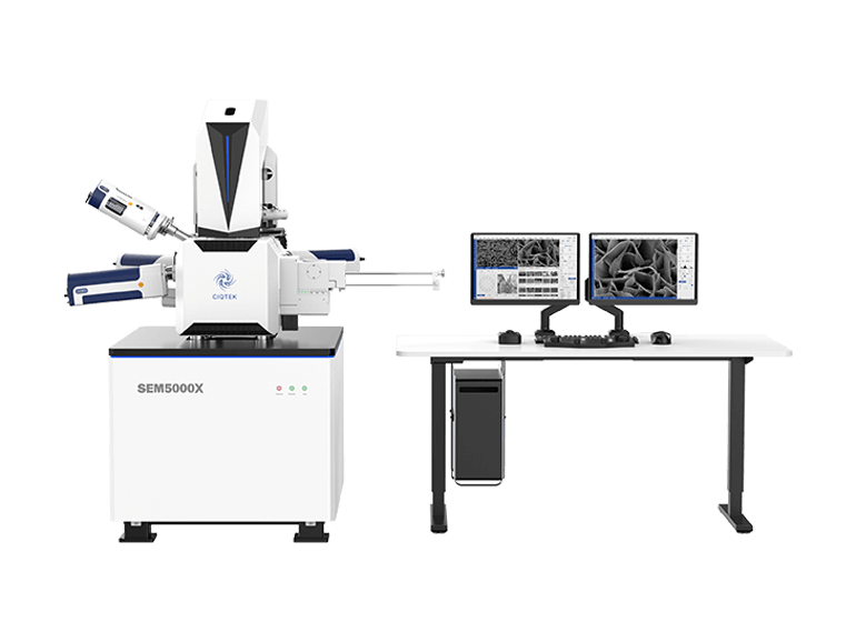
Nuclear fusion is considered a key future energy source due to its high efficiency and clean energy output. In fusion reactors, water cooling systems are widely used because they are technically mature, cost-effective, and have excellent cooling performance. However, a major challenge remains: under high temperature and high pressure, water and steam strongly corrode structural materials. While this problem has been studied in fission reactors, fusion environments are more complex. The unique high-intensity, unevenly distributed magnetic fields in fusion devices interact with corrosion processes, creating new technical challenges that need detailed research. To address this, Associate Professor Peng Lei's team from the University of Science and Technology of China conducted an in-depth study using the CIQTEK scanning electron microscope (SEM) and dual-beam electron microscope. They built high-temperature magnetic-field steam corrosion and high-temperature water corrosion setups. Using SEM, EBSD, and FIB techniques, they analyzed oxide films formed on CLF-1 steel after 0–300 hours of steam corrosion at 400°C under 0T, 0.28T, and 0.46T magnetic fields, and after 1000 hours of high-temperature water corrosion at 300°C.   The study used CIQTEK SEM5000X ultra-high-resolution field-emission SEM and the FIB-SEM DB500   The study found that the oxide films form a multilayer structure, with a chromium-rich inner layer and an iron-rich outer layer. Film formation occurs in five stages: initial oxide particles, then floc-like structures, formation of a dense layer, growth of spinel structures on the dense layer, and finally, spinel cracking into laminated oxides. The presence of a magnetic field significantly accelerates corrosion, promotes the transformation of outer magnetite (Fe₃O₄) into hematite (Fe₂O₃), and enhances laminated oxide formation. This work was published in Corrosion Science, a top-tier journal in the field of corrosion and materials degradation, under the title: "Magnetic field effects on the high-temperature steam corrosion behavior of reduced activation ferritic/martensitic steel."     Surface Oxide Film Characterization In high-temperature steam (HTS), CLF-1 steel surfaces show different corrosion states over time. On polished surfaces, early-stage oxidation (60 h) appears as small, dispersed particles. The Fe/Cr ratio is similar to the substrate, indicating that the oxide layer is not yet complete. By 120 h, floc-like oxides appear. At 200 h, a dense oxide layer forms, with new oxide particles and local spinel structures on top. Rough surfaces corrode faster. Early floc-like oxides are finer and more evenly distributed. By 200 h, they transform into spinel structures, showing a stronger difference from polished surfaces. In high-temperature, high-pressure water (HTPW), polished surfaces display similar spinel structures. Spinel in HTPW is denser and more numerous, while spinel in HTS is larger in size....
CIQTEK SEM and EPR Reveal a New Pathway for Nickel-Citrate Removal
With the rapid expansion of new energy, mining, metallurgy, and electroplating industries, nickel pollution in water bodies has become a growing threat to environmental quality and human health. During industrial processes, nickel ions often interact with various chemical additives to form highly stable heavy-metal organic complexes (HMCs). In nickel electroplating, for example, citrate (Cit) is widely used to improve coating uniformity and brightness, but the two carboxyl groups in Cit readily coordinate with Ni²⁺ to form Ni–Citrate (Ni-Cit) complexes (logβ = 6.86). These complexes significantly alter nickel’s charge, steric configuration, mobility, and ecological risks, while their stability makes them challenging to remove with conventional precipitation or adsorption methods. Currently, "complex dissociation" is regarded as the key step in removing HMCs. However, typical oxidation or chemical treatments suffer from high cost and complicated operation. Therefore, multifunctional materials with both oxidative and adsorptive capabilities offer a promising alternative. Researchers from Beihang University, led by Prof. Xiaomin Li and Prof. Wenhong Fan, used the CIQTEK scanning electron microscope (SEM) and electron paramagnetic resonance (EPR) spectrometer to conduct an in-depth investigation. They developed a new strategy using KOH-modified Arundo donax L. biochar to efficiently remove Ni-Cit from water. The modified biochar not only showed high removal efficiency but also enabled nickel recovery on the biochar surface. The study, titled “Removal of Nickel-Citrate by KOH-Modified Arundo donax L. Biochar: Critical Role of Persistent Free Radicals”, was recently published in Water Research.     Material Characterization Biochar was produced from Arundo donax leaves and impregnated with KOH at different mass ratios. SEM imaging (Fig. 1) revealed: The original biochar (BC) exhibited a disordered rod-like morphology. At a 1:1 KOH-to-biomass ratio (1KBC), an ordered honeycomb-like porous structure was formed. At ratios of 0.5:1 or 1.5:1, pores were underdeveloped or collapsed. BET analysis confirmed the highest surface area for 1KBC (574.2 m²/g), far exceeding other samples. SEM and BET characterization provided clear evidence that KOH modification dramatically enhances porosity and surface area—key factors for adsorption and redox reactivity.   Figure 1. Preparation and characterization of KOH-modified biochar.   Performance in Ni-Cit Removal Figure 2. (a) Removal efficiency of total Ni by different biochars; (b) TOC variation during Ni–Cit treatment; (c) Effect of Ni–Cit concentration on the removal efficiency of 1KBC; (d) Effect of pH on the removal performance of 1KBC; (e) Influence of coexisting ions on Ni–Cit removal by 1KBC; (f) Continuous-flow removal performance of Ni–Cit by 1KBC. (Ni–Cit = 50 mg/L, biochar dosage = 1 g/L)   Batch experiments de...
Top

Leave A Message

Leave A Message
Please feel free to contact us for more details, request a quote or book an online demo! We will reply you as soon as we can.
Submit

Home

Products

Chat

contact