Products

Products

CIQTEK is the manufacturer and global supplier of high-performance scientific instruments, such as Electron Microscopes, Electron Paramagnetic Resonance (Electron Spin Resonance), Gas Adsorption Analyzers, Scanning NV Microscopes, etc.
Applications
Exploring Rice - Scanning Electron Microscope (SEM) Applications
To begin with, what is aged rice and new rice? Aged rice or old rice is nothing but stocked rice that is kept for aging for one or more years. On the other hand, new rice is the one which is produced from newly harvested crops. Compared to the fresh aroma of new rice, aged rice is light and tasteless, which is essentially a change in the internal microscopic morphological structure of aged rice. Researchers analyzed new rice and aged rice using the CIQTEK tungsten filament scanning electron microscope SEM3100. Let's see how they differ in the microscopic world!   CIQTEK Tungsten Filament Scanning Electron Microscope SEM3100   Figure 1 Cross-sectional fracture morphology of new rice and aged rice   First, the microstructure of rice endosperm was observed by SEM3100. From Figure 1, it can be seen that the endosperm cells of new rice were long polygonal prismatic cells with starch grains wrapped in them, and the endosperm cells were arranged in a radial fan shape with the center of the endosperm as concentric circles, and the endosperm cells in the center were smaller compared with the outer cells. The radial fan-shaped endosperm structure of new rice was more obvious than that of aged rice.   Figure 2 Microstructure morphology of the central endosperm of new rice and aged rice   Further magnified observation of the central endosperm tissue of rice revealed that the endosperm cells in the central part of aged rice were more broken and the starch granules were more exposed, making the endosperm cells radially arranged in a blurred form.   Figure 3 Microstructure morphology of protein film on the surface of new rice and aged rice   The protein film on the surface of the endosperm cells was observed at high magnification using the advantages of SEM3100 with high-resolution imaging. As can be seen from Figure 3, a protein film could be observed on the surface of new rice, while the protein film on the surface of aged rice was broken and had different degrees of warping, resulting in relatively clear exposure of the internal starch granule shape due to the reduction of the surface protein film thickness.    Figure 4 Microstructure of endosperm starch granules of new rice   Rice endosperm cells contain single and compound amyloplasts. Single-grain amyloplasts are crystalline polyhedra, often in the form of single grains with blunt angles and obvious gaps with the surrounding amyloplasts, containing mainly crystalline and amorphous regions formed by straight-chain and branched-chain amylose [1,2]. The complex grain amyloplasts are angular in shape, densely arranged, and tightly bound to the surrounding amyloplasts. Studies have shown that the starch grains of high-quality rice exist mainly as complex grains [3]. By observing the endosperm cells of new rice, as shown in Figure 4, the starch grains mostly existed in the form of compound grains. The compound starch grains were angular in shape and closely...
Electronic Ceramics Analysis - Scanning Electron Microscopy (SEM) Applications
Ceramic materials have a series of characteristics such as high melting point, high hardness, high wear resistance, and oxidation resistance, and are widely used in various fields of national economy such as the electronics industry, automotive industry, textile, chemical industry, and aerospace. The physical properties of ceramic materials depend largely on their microstructure, which is an important application area of SEM.     What are ceramics? Ceramic materials are a class of inorganic non-metallic materials made of natural or synthetic compounds through forming and high-temperature sintering and can be divided into general ceramic materials and special ceramic materials.   Special ceramic materials can be classified according to chemical composition: oxide ceramics, nitride ceramics, carbide ceramics, boride ceramics, silicide ceramics, etc.; according to their characteristics and applications can be divided into structural ceramics and functional ceramics.   Figure 1 Microscopic morphology of boron nitride ceramics   SEM helps to study the properties of ceramic materials   With the continuous development of society and science and technology, people's requirements for materials have been increasing, which requires a deeper understanding of the various physical and chemical properties of ceramics. The physical properties of ceramic materials are largely dependent on their microstructure [1], and SEM images are widely used in ceramic materials and other research fields because of their high resolution, wide adjustable magnification range, and stereoscopic imaging. The CIQTEK Field Emission Scanning Electron Microscope SEM5000 can be used to observe the microstructure of ceramic materials and related products easily, and in addition, the X-ray energy spectrometer can be used to determine the elemental composition of materials quickly.    Application of SEM in the Study of Electronic CeramicsThe largest end-use market of the special ceramics industry is the electronics industry, where barium titanate (BaTiO3) is widely used in multilayer ceramic capacitors (MLCC), thermistors (PTC), and other electronic components because of its high dielectric constant, excellent ferroelectric and piezoelectric properties, and voltage resistance and insulation properties [2]. With the rapid development of the electronic information industry, the demand for barium titanate is increasing, and the electronic components are becoming smaller and more miniaturized, which also puts forward higher requirements for barium titanate.Researchers often regulate the properties by changing the sintering temperature, atmosphere, doping, and other preparation processes. Still, the essence is that the changes in the preparation process cause changes in the microstructure of the material and thus the properties. Studies have shown that the dielectric ferroelectric properties of barium titanate are closely related to the material's mi...
Explore Pollen Micromorphology - Scanning Electron Microscope (SEM) Applications
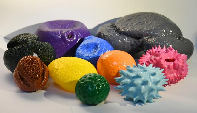
In scientific research, pollen has a wide range of applications. According to Dr. Limi Mao, Nanjing Institute of Geology and Paleontology, Chinese Academy of Sciences, by extracting and analyzing different pollen deposited in the soil, it is possible to understand which parent plants they came from respectively, and thus infer the environment and climate at that time. In the field of botanical research, pollen mainly provides microscopic reference evidence for systematic taxonomy. More interestingly, pollen evidence can also be applied in criminal investigation cases. Forensic palynology can effectively corroborate the facts of a crime by using pollen spectrum evidence on the suspect's accompanying clothing and at the crime scene. In the field of geological research, pollen has been widely used in reconstructing vegetation history, past ecology, and climate change studies. In archaeological studies exploring early human farming civilizations and habitats, pollen can help scientists understand the history of early human domestication of plants, what food crops were cultivated, etc.    Fig. 1 3D pollen model picture (taken by Dr. Limi Mao, product developed by Dr. Oliver Wilson)   The size of pollen varies from a few microns to more than two hundred microns, which is beyond the resolution of visual observation and requires the use of a microscope for observation and study. Pollen comes in a wide variety of morphologies, including variations in size, shape, wall structure, and ornamentation. The ornamentation of pollen is one of the key bases for identifying and distinguishing pollen. However, the resolution of the optical biological microscope has physical limitations, it is difficult to precisely observe the differences between different pollen ornamentation, and even the ornamentation of some small pollen cannot be observed. Therefore, scientists need to use a scanning electron microscope (SEM) with high resolution and large depth of field to obtain a clear picture of pollen morphological features. In the study of fossil pollen, it is possible to identify the specific plants to which the pollen belongs, so as to more accurately understand the vegetation, environment, and climate information of the time.     The Microstructure of Pollen   Recently, researchers have used the CIQTEK Tungsten Filament SEM3100 and the CIQTEK Field Emission SEM5000 to microscopically observe a variety of pollen.  Fig. 2 CIQTEK Tungsten Filament SEM3100 and Field Emission SEM5000   1. Cherry blossom Pollen grains spherical-oblong. With three pore grooves (without treated pollen, the pores are not obvious), the grooves reach both poles. Outer wall with striate ornamentation.     2. Chinese violet cress (Orychophragmus violaceus) Chinese violet cress pollen morphology is ellipsoidal, with 3 grooves, the surface has a reticulated pattern, and the mesh size varies.     3. Ottelia Pollen grains are rounded, wit...
Learn More
Leave A Message
Submit
Top

Leave A Message

Leave A Message
Please feel free to contact us for more details, request a quote or book an online demo! We will reply you as soon as we can.
Submit

Home

Products

Chat

contact