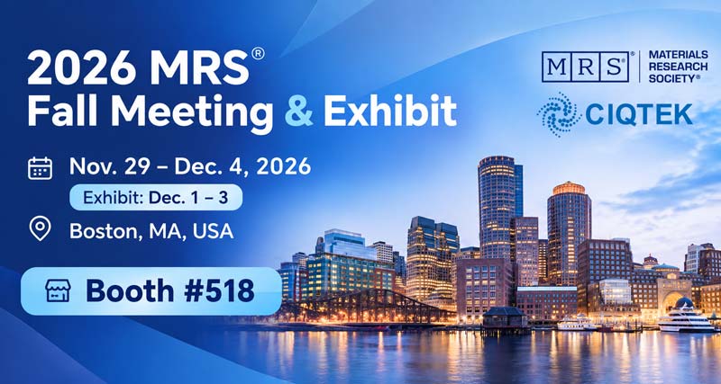
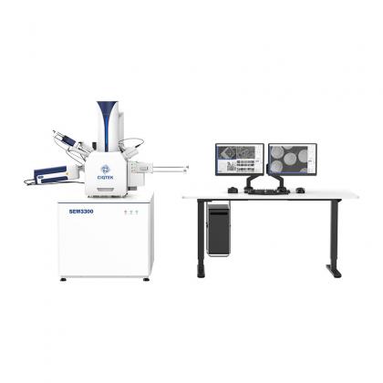
CIQTEK is heading to Boston for the 2026 MRS Fall Meeting & Exhibit, taking place from November 29 to December 4, 2026. Our U.S. electron microscopy team will be on-site at Booth #518, ready to meet researchers working across materials science, microscopy, and advanced characterization. This year, CIQTEK will also take part in the scientific program with a presentation focused on HEM, our High-Speed SEM technology, and how faster electron imaging can support more efficient microscopy workflows. Join Our HEM Presentation Session: CH05.05: Applied AI in Electron Microscopy II Date: December 2, 2026 Session Time: 8:00 AM–12:00 PM ET CIQTEK Presentation: 9:15 AM–9:30 AM ET Speaker: Xin Ren, Senior Electron Microscopy Engineer, CIQTEK The presentation will explore high-speed electron microscopy and the role of HEM in accelerating image acquisition while supporting demanding materials characterization workflows. Meet Us at Booth #518 If you are attending MRS Fall 2026, stop by Booth #518 to meet the CIQTEK U.S. team and talk about your SEM, FIB-SEM, and electron microscopy applications. Whether you are working on advanced materials, semiconductor research, or new characterization workflows, we would be glad to hear what you are working on and discuss how CIQTEK electron microscopy solutions may fit your research. See you in Boston!
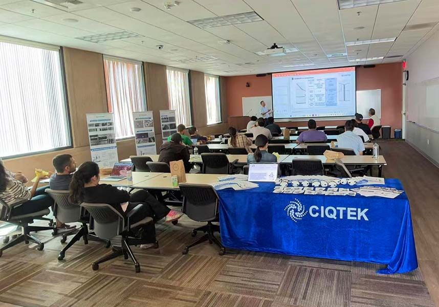
View MoreOn November 3, 2026, CIQTEK hosted an electron microscopy seminar at a university in California, bringing researchers together for a practical discussion on SEM technology, FIB, high-speed imaging, and the future of integrated characterization. The seminar brought together researchers and microscopy users, with lively discussion around SEM technology, applications, and future workflows. Rather than focusing only on instrument specifications, the conversation explored how microscopy is evolving around real research needs and more efficient ways of working. From SEM Imaging to Integrated Characterization One of the main themes was the changing role of the scanning electron microscope. SEM is no longer only an imaging tool. New workflows increasingly combine high-resolution imaging, sample preparation, analysis, and other characterization methods into a more connected platform. The seminar also explored how FIB and SEM technologies can work together in applications that require site-specific preparation, cross-sectioning, and deeper structural analysis. For researchers, the focus is on how these technologies can support real applications and deliver practical value in microscopy workflows. Faster Imaging with High-Speed SEM Another topic was HEM, or High-Speed SEM, one of CIQTEK's recent developments in electron microscopy. The technology is designed to increase image acquisition speed while maintaining high-resolution imaging. CIQTEK shared how faster acquisition can support workflows where large amounts of image data need to be collected efficiently. This can be especially useful in applications involving large-area imaging, repeated measurements, or high-throughput characterization. Continuing the Conversation For CIQTEK, events like this are not only a way to introduce new technology. They are also a chance to hear directly from researchers and better understand how electron microscopy is being used in real laboratories. CIQTEK plans to continue organizing more technical seminars and research exchanges across the United States. If you are interested in hosting a similar seminar or learning more about CIQTEK electron microscopy solutions, feel free to contact our U.S. team.
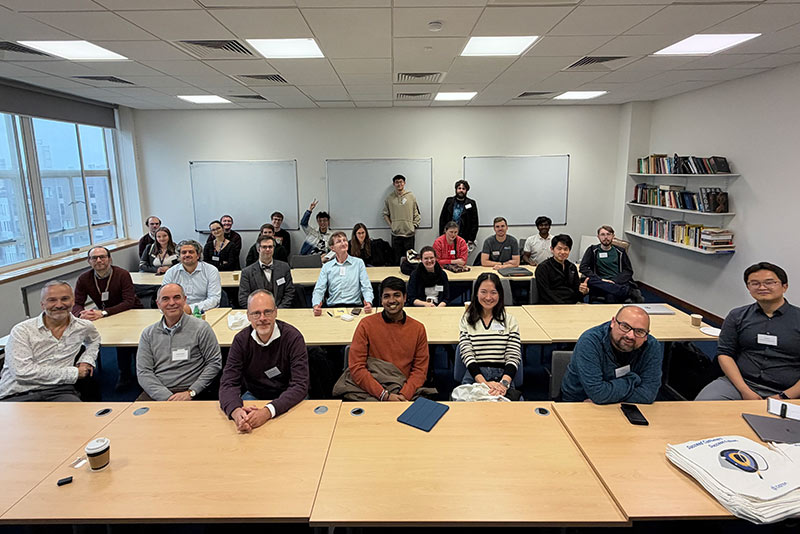
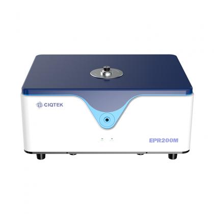
View MoreOn October 21, CIQTEK and Queen Mary University of London brought together EPR researchers and specialists from leading European universities for a full day of scientific exchange in London. The programme covered a wide range of topics, from molecular actinides and biometalloclusters to molecular spin systems and next-generation EPR instrumentation. Different Research Questions, One EPR Community The talks showed just how broad modern EPR spectroscopy has become. Researchers from Queen Mary University of London, The University of Manchester, University of Oxford, University of Stuttgart, University of Copenhagen, and CIQTEK shared how EPR is being used to answer very different scientific questions. That variety made the day especially useful. Attendees could compare approaches across chemistry, physics, molecular science, and magnetic resonance rather than looking at EPR from only one angle. The Conversation Continued Around the Instrument After the presentations, the discussion moved from slides to the instrument itself. Attendees gathered around a CIQTEK floor-standing EPR spectrometer on site, continuing the conversation around real hardware, system configuration, experimental workflows, and practical research needs. Questions quickly became more specific: How would a certain experiment be set up? Which configuration would fit a particular research problem? What should researchers consider when moving from an idea to a real EPR measurement? That exchange was one of the most valuable parts of the day. More Conversations to Come By combining scientific talks with direct discussion around the instrument, the workshop created a practical space for researchers to exchange ideas, ask questions, and explore how EPR can support their work. Thank you to everyone who joined us at Queen Mary University of London and helped make the day such a meaningful exchange. CIQTEK plans to organize more EPR seminars, workshops, and scientific exchange events across Europe and the United States in the future. If you are interested in attending, speaking, or collaborating on a similar event, we would be happy to hear from you. Contact us to stay connected and learn about upcoming EPR events!
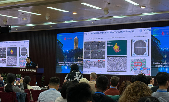
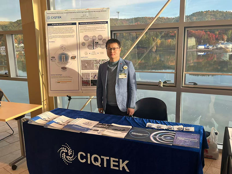
View MoreCIQTEK has wrapped up its participation in the 2026 vEM Technology Forum at Peking University in Beijing, where researchers and microscopy specialists gathered to discuss the latest developments in Volume Electron Microscopy, large-area imaging, automation, and 3D reconstruction. For CIQTEK, the conversations came back to one practical challenge again and again: how to image larger biological volumes faster without giving up the resolution needed to see fine structures. That is exactly where high-speed SEM is becoming increasingly important. From Faster Imaging to Larger Volume EM Workflows At the CIQTEK booth, our team introduced the HEM6000 High-Speed SEM and discussed how high imaging speed, low-kV resolution, automated large-area acquisition, and intelligent workflows can support increasingly data-intensive Volume EM research. For researchers working with large numbers of serial sections or extended regions of interest, acquisition speed is only one part of the workflow. Stable imaging, consistent quality across large areas, and automation become equally important as datasets grow. These topics led to many useful discussions with researchers working on biological imaging, neuroscience, connectomics, and other large-scale microscopy applications. Sharing Our Latest Work on High-Speed SEM During the forum, Dr. Changming Liu (Noah), CIQTEK Senior Electron Microscopy Solution Manager, presented CIQTEK's latest work on high-speed SEM for large-scale Volume EM. The presentation highlighted how the HEM6000 combines high-speed acquisition with low-kV imaging performance, large-area imaging, and automation to make demanding Volume EM workflows more efficient. Rather than treating speed as a standalone specification, the discussion focused on a broader question: how can faster acquisition translate into a more practical workflow for real research? More Conversations Ahead The 2026 vEM Technology Forum was a valuable opportunity to hear directly from the microscopy community about the challenges behind large-scale imaging and data acquisition. Those conversations also give us a clearer view of where electron microscopy workflows are heading: larger datasets, more automation, higher throughput, and stronger demand for reliable high-resolution imaging at scale. CIQTEK will continue developing electron microscopy solutions that help researchers move from individual high-resolution images toward more efficient, large-scale characterization workflows. Thank you to everyone who stopped by, shared ideas, and discussed their research with us in Beijing.
View MoreCIQTEK will join Neuroscience 2026, the annual meeting of the Society for Neuroscience, in Washington, D.C. from November 14 to 18. Visit us at Booth #1014 to explore how high-speed electron microscopy can support large-scale biological imaging, connectomics, and volume electron microscopy workflows. Neuroscience 2026 will take place at the Walter E. Washington Convention Center and brings together neuroscientists, imaging specialists, technology providers, and researchers from around the world. High-Speed SEM for Large-Scale Biological Imaging Modern neuroscience increasingly depends on imaging larger areas, larger volumes, and more complex biological structures. For volume electron microscopy, imaging speed matters, but speed alone is not enough. Researchers also need stable low-kV performance, reliable image quality, automated acquisition, and workflows that can handle large datasets efficiently. At Neuroscience 2026, CIQTEK will highlight the HEM6000 High-Speed SEM, developed for demanding applications in biological imaging and large-area electron microscopy. Its capabilities support workflows such as: High-speed large-area imaging Low-kV high-resolution observation Automated image acquisition Serial section imaging Volume EM and 3D reconstruction Neuroscience and connectomics research The goal is to help researchers collect high-quality structural information faster while reducing the time and effort required for large-scale imaging projects. Explore CIQTEK Electron Microscopy Solutions In addition to high-speed SEM, CIQTEK offers a broader electron microscopy portfolio for biological and materials research, including field emission SEM and FIB-SEM systems. Our team will be available to discuss practical questions around sample imaging, throughput, resolution, automation, and workflow design. If you are working with brain tissue, neural structures, connectomics, biological ultrastructure, or large-volume imaging, we would be glad to learn more about your research and discuss which electron microscopy approach may fit your needs. Date: November 14–18, 2026Location: Walter E. Washington Convention Center, Washington, D.C., USACIQTEK Booth: #1014 Meet the CIQTEK team at Booth #1014 and see how faster electron microscopy can support your next neuroscience imaging project.
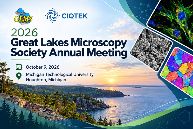
View MoreSometimes the most useful microscopy conversations happen away from a formal presentation, with a sample, an image, or a practical question on the table. That was our experience at the 2026 Great Lakes Microscopy Society Annual Meeting, held on October 9 at Michigan Technological University in Houghton, Michigan. CIQTEK joined researchers, students, and microscopy professionals working across electron microscopy, microanalysis, materials science, and biological research. Microscopy Questions, Not Just Instrument Specs Throughout the meeting, our U.S. Electron Microscopy Team had the chance to talk with attendees about what they actually need from an SEM workflow, from routine imaging and materials characterization to higher-resolution and more specialized applications. CIQTEK also shared its broader electron microscopy portfolio, covering field emission SEM, tungsten SEM, high-speed SEM, and FIB-SEM solutions for different research needs. Good Science Starts with Good Conversations GLMS is a smaller, focused meeting, and that made it especially easy to have direct conversations with people doing hands-on microscopy every day. Thank you to everyone who stopped by, asked questions, shared your research, or simply came over for a microscopy chat. For our team, meetings like GLMS are valuable because they help us understand how researchers are actually using electron microscopy, where the challenges are, and what they expect from the next generation of instruments. We look forward to staying connected with the Great Lakes microscopy community and continuing the conversations started in Michigan.
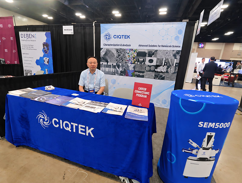
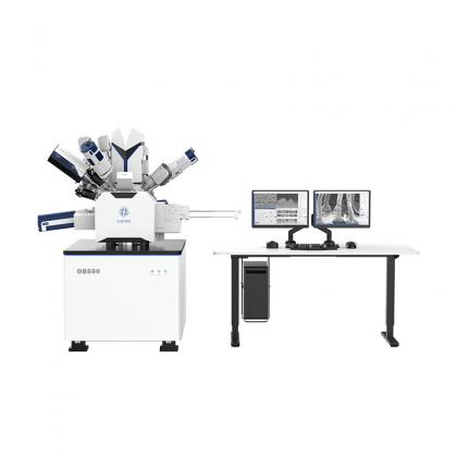
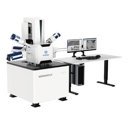
View MoreFailure analysis rarely starts with a perfect sample. At ISTFA 2026, the 52nd International Symposium for Testing and Failure Analysis, many of our conversations began with real-world challenges: buried defects, difficult cross sections, subtle material contrast, and structures that require more than surface-level imaging. From October 4 to 8 in San Antonio, Texas, CIQTEK U.S. Electron Microscopy Team met with failure analysis engineers, semiconductor researchers, and microscopy specialists at Booth #317 to discuss practical SEM and FIB-SEM workflows. From Surface Imaging to Buried Defect Analysis Discussions at ISTFA covered a wide range of applications, including: Semiconductor device and wafer inspection High-resolution low-kV imaging Surface defect and morphology analysis Material contrast and contamination analysis Site-specific cross-sectioning Buried defect and interface characterization SEM helps reveal surface morphology, interfaces, particles, and material contrast, while FIB-SEM extends the analysis beneath the surface through precise milling and targeted cross-section preparation. CIQTEK also introduced its electron microscopy solutions, including the SEM5000 Series and DB550 FIB-SEM, for semiconductor failure analysis and advanced materials characterization. Valuable Conversations at Booth #317 Beyond instrument specifications, many discussions focused on real laboratory needs such as sample type, imaging conditions, detector configuration, workflow efficiency, and cross-section quality. These conversations gave our team valuable insight into the challenges faced by semiconductor and failure analysis laboratories in the U.S. Thank you to everyone who visited our booth and shared your applications and ideas with us. We look forward to continuing the conversation and supporting more laboratories with practical electron microscopy solutions for failure analysis and defect characterization.
View MoreCIQTEK will participate in the 2026 Great Lakes Microscopy Society (GLMS) Annual Meeting on October 9, 2026, at Michigan Technological University in Houghton, Michigan. The meeting brings together researchers, students, and microscopy professionals working across electron microscopy, microanalysis, materials science, and biological research. Meet the CIQTEK U.S. EM Team At GLMS 2026, CIQTEK will showcase its electron microscopy solutions for a wide range of research applications. Our portfolio includes field emission SEM, tungsten SEM, high-speed SEM, and FIB-SEM. Our U.S. Electron Microscopy Team will be on-site to connect with attendees and discuss real microscopy applications and workflow needs. Experienced EM Solution Managers will also be available to answer technical questions and help researchers explore suitable microscopy solutions for their labs. Date: October 9, 2026Location: Michigan Technological University, Houghton, Michigan, USA See you at GLMS 2026!
View More
No. 1969, Kongquetai Road, High-tech Zone, Hefei, Anhui, China
info@ciqtek.com
Sitemap | XML | Blog | Privacy Policy | IPv6 network supported