CIQTEK to Join Neuroscience 2026 with Volume EM Solutions, USA
October 16, 2026
CIQTEK will join Neuroscience 2026, the annual meeting of the Society for Neuroscience, in Washington, D.C. from November 14 to 18. Visit us at Booth #1014 to explore how high-speed electron microscopy can support large-scale biological imaging, connectomics, and volume electron microscopy workflows.
Neuroscience 2026 will take place at the Walter E. Washington Convention Center and brings together neuroscientists, imaging specialists, technology providers, and researchers from around the world.
High-Speed SEM for Large-Scale Biological Imaging
Modern neuroscience increasingly depends on imaging larger areas, larger volumes, and more complex biological structures.
For volume electron microscopy, imaging speed matters, but speed alone is not enough. Researchers also need stable low-kV performance, reliable image quality, automated acquisition, and workflows that can handle large datasets efficiently.
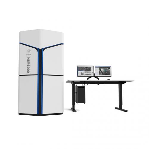
At Neuroscience 2026, CIQTEK will highlight the HEM6000 High-Speed SEM, developed for demanding applications in biological imaging and large-area electron microscopy.
Its capabilities support workflows such as:
High-speed large-area imaging
Low-kV high-resolution observation
Automated image acquisition
Serial section imaging
Volume EM and 3D reconstruction
Neuroscience and connectomics research
The goal is to help researchers collect high-quality structural information faster while reducing the time and effort required for large-scale imaging projects.
Explore CIQTEK Electron Microscopy Solutions
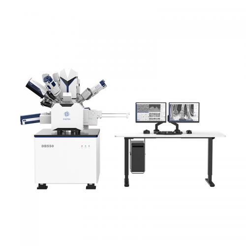
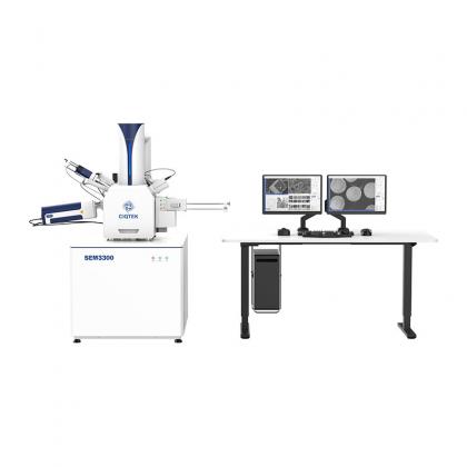
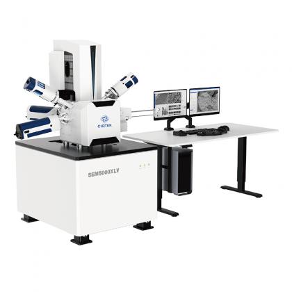
In addition to high-speed SEM, CIQTEK offers a broader electron microscopy portfolio for biological and materials research, including field emission SEM and FIB-SEM systems.
Our team will be available to discuss practical questions around sample imaging, throughput, resolution, automation, and workflow design.
If you are working with brain tissue, neural structures, connectomics, biological ultrastructure, or large-volume imaging, we would be glad to learn more about your research and discuss which electron microscopy approach may fit your needs.
Date: November 14–18, 2026 Location: Walter E. Washington Convention Center, Washington, D.C., USA CIQTEK Booth: #1014
Meet the CIQTEK team at Booth #1014 and see how faster electron microscopy can support your next neuroscience imaging project.
High-speed Fully Automated Field Emission Scanning Electron Microscope Workstation CIQTEK HEM6000 facilities technologies such as the high-brightness large-beam current electron gun, high-speed electron beam deflection system, high-voltage sample stage deceleration, dynamic optical axis, and immersion electromagnetic & electrostatic combo objective lens to achieve high-speed image acquisition whilst ensuring nano-scale resolution. The automated operation process is designed for applications such as a more efficient and smarter large-area high-resolution imaging workflow. Its imaging speed is over five times faster than that of a conventional field emission scanning electron microscope (FESEM).
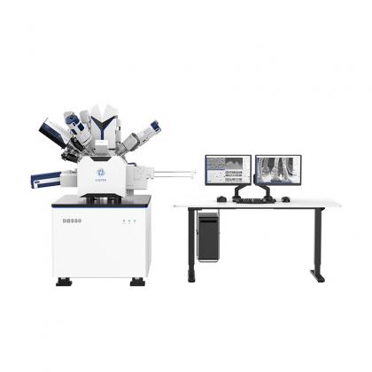
Ga+ Focused Ion Beam Field Emission Scanning Electron Microscope The CIQTEK DB550 Focused Ion Beam Scanning Electron Microscope (FIB-SEM) has a focused ion beam column for nano-analysis and specimen preparation. It utilizes “super tunnel” electron optics technology, low aberration, and non-magnetic objective design, and has the “low voltage, high resolution” feature to ensure its nanoscale analytical capabilities. The ion columns facilitate a Ga+ liquid metal ion source with highly stable and high-quality ion beams to ensure nanofabrication capabilities. The DB550 is an all-in-one nano-analysis and fabrication workstation with an integrated nano-manipulator, gas injection system, and user-friendly GUI software.